Welcome to the Amira-Avizo Software Use Case Gallery
Below you will find a collection of use cases of our 3D data visualization and analysis software. These use cases include scientific publications, articles, papers, posters, presentations or even videos that show how Amira-Avizo Software is used to address various scientific and industrial research topics.
Use the Domain selector to filter by main application area, and use the Search box to enter keywords related to specific topics you are interested in.
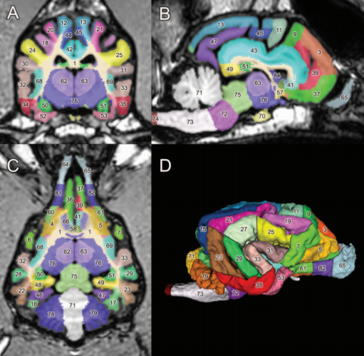
Classification and delineation of the motor-related nuclei in the human thalamus have been the focus of numerous discussions for a long time. Difficulties in finding consensus have for the most part been caused by paucity of direct experimental data on connections of individual nuclear entities. Kultas-Ilinsky et al. (2011, J Comp Neurol, 519:2811-2837) showed that distribution of the isoform 65 of glutamic acid decarboxylase (GAD65), the enzyme that synthesizes inhibitory neurotransmitt... Read more
Igor Ilinsky, Andreas Horn, Perrine Paul-Gilloteaux, Pierre Gressens, Catherine Verney, Kristy Kultas-Ilinsky
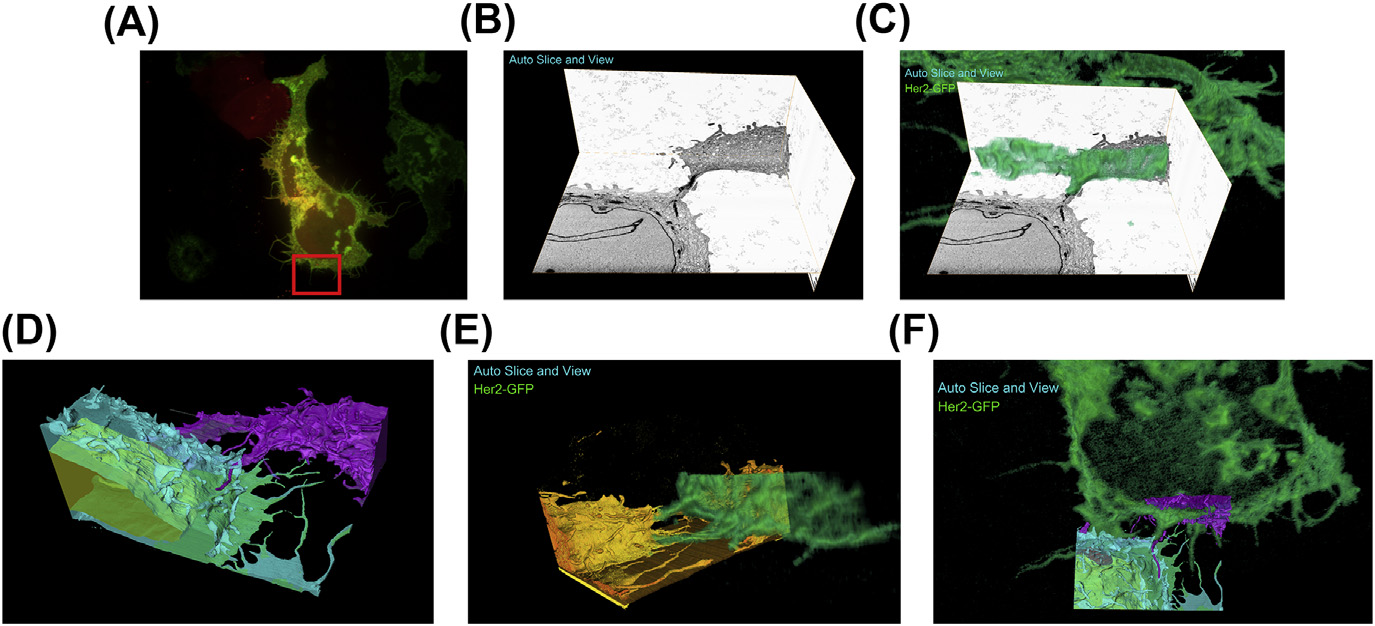
A fully integrated, three-dimensional fluorescence to electron microscopy correlative workflow
While fluorescence microscopy provides tools for highly specific labeling and sensitive detection, its resolution limit and lack of general contrast has hindered studies of cellular structure and protein localization. Recent advances in correlative light and electron microscopy (CLEM), including the fully integrated CLEM workflow instrument, the Thermo Scientific CorrSight with MAPS, have allowed for a more reliable, reproducible, and quicker approach to correlate three-dimensional time-lapse... Read more
Claudia S. Lopez, Cedric Bouchet-Marquis, Christopher P. Arthur, Jessica L. Riesterer, Gregor Heiss, Guillaume Thibault, Lee Pullan, Sunjong Kwon, Joe W. Gray
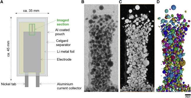
Nickel-rich transition metal oxide materials […] are of great interest for achieving immediate improvements in the energy density of Li-ion batteries and for risk reduction within the Li-ion battery supply chain.
[…] An increase in Ni content in NMC materials leads to accelerated degradation […]. This potentially complicates their adoption in applications requiring extended cycle life such as in electric vehicles.
Recent developments in X-ray characterization to... Read more
ChunTan, Andrew S.Leach, Thomas M.M. Heenan, Huw Parks, Rhodri Jervis, Johanna Nelson Weker, Daniel J.L. Brett, Paul R.Shearing
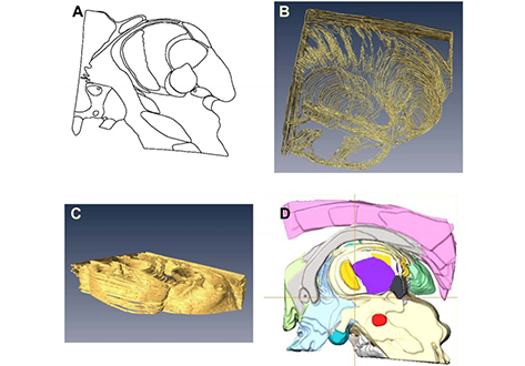
A detailed canine brain label map for neuroimaging analysis
Dogs have recently become an important model species for comparative social and cognitive neuroscience. Brain template-related label maps are essential for functional magnetic resonance imaging (fMRI) data analysis, to localize neural responses. In this study, we present a detailed, individual-based, T1-weighted MRI-based brain label map used in dog neuroimaging analysis. Methods: A typical, medium-headed dog (a 7.5-year-old
male Golden Retriever) was selected from a cohort of ... Read more
Czeibert Kálmán, Andics Attila, Petneházy Örs, Kubinyi Enikő, Kálmán Czeibert
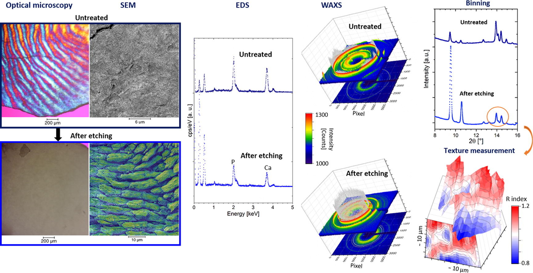
Enamel caries is a highly prevalent worldwide disease that involves the demineralisation of the outer tooth structure. In this study, we report the analysis of artificially demineralised human enamel sections (‘slices’) etched using lactic acid (2% v/v) in comparison with healthy enamel using correlative techniques of optical and electron microscopy, as well as scanning diffraction. Demineralisation of the enamel was characterised at the micron to sub-micron scale. The structure of the he... Read more
Cyril Besnard, Robert A. Harper, Thomas E. J. Moxham, Jonathan D. James, Malte Storm, Enrico Salvati, Gabriel Landini, Richard M. Shelton, Alexander M.Korsunsky
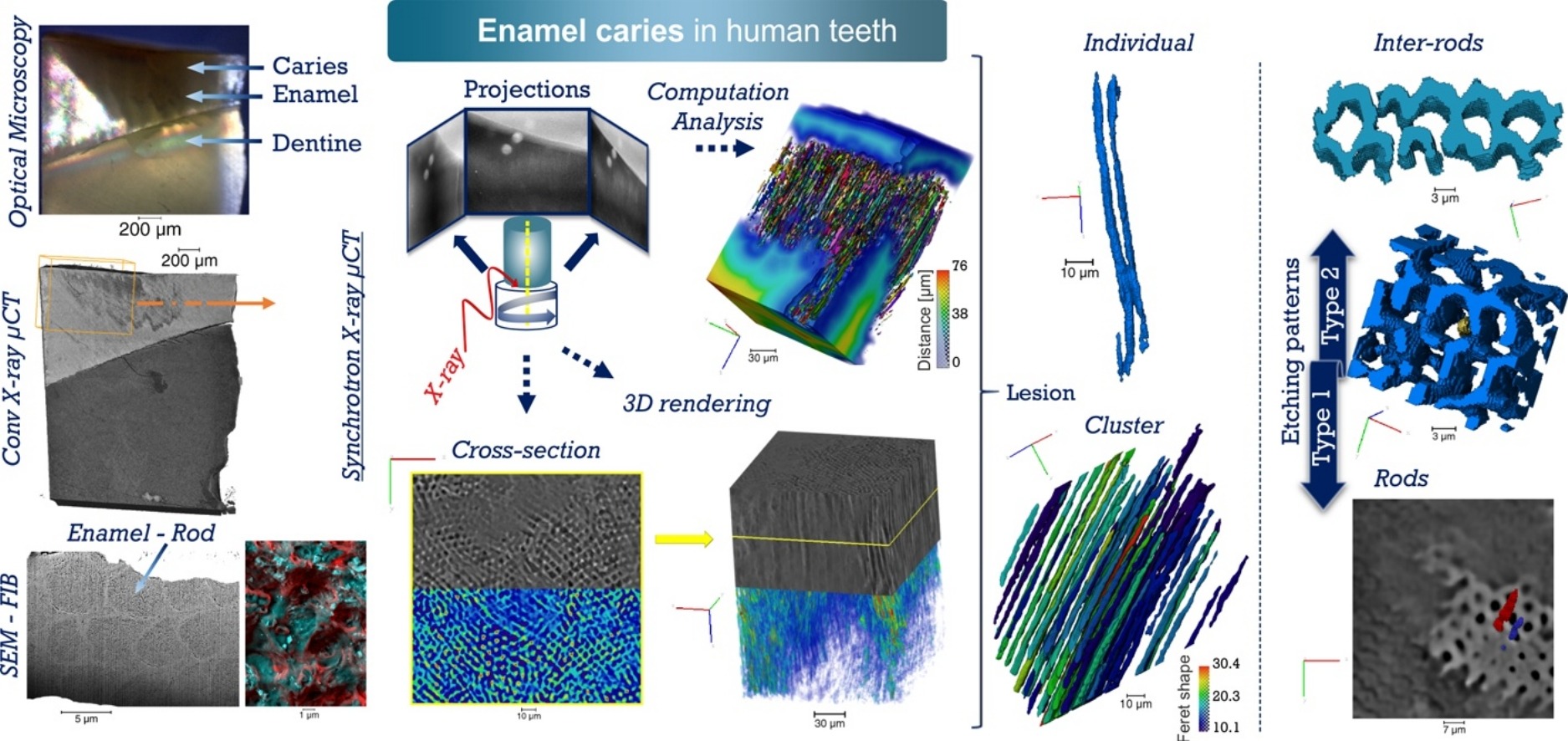
Unprecedented combination of resolution, field of view and contrast for the analysis human enamel carious lesions was achieved. Synchrotron X-ray micro-computed tomography revealed sub-micron details of enamel rod and inter-rod regions inaccessible by laboratory tomography. Successful segmentation and labelling allowed the extraction of enamel etching patterns and statistics. Correlation was obtained between synchrotron X-ray micro-tomography and FIB-SEM cross-sec... Read more
Cyril Besnard, Robert A. Harper, Thomas E. J. Moxham, Jonathan D. James, Malte Storm, Enrico Salvati, Gabriel Landini, Richard M. Shelton, Alexander M.Korsunsky
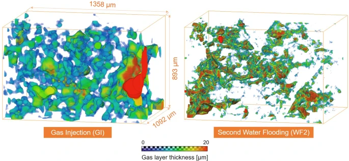
Pore-scale mechanisms of CO2 storage in oilfields
Rapid implementation of global scale carbon capture and storage is required to limit temperature rises to 1.5 °C this century. Depleted oilfields provide an immediate option for storage, since injection infrastructure is in place and there is an economic benefit from enhanced oil recovery. To design secure storage, we need to understand how the fluids are configured in the microscopic pore spaces of the reservoir rock. We use high-resolution X-ray imaging to study the flow of oil, water and ... Read more
Abdulla Alhosani, Alessio Scanziani, Qingyang Lin, Ali Q. Raeini, Branko Bijeljic & Martin J. Blunt

Defect structure process maps for laser powder bed fusion additive manufacturing
Accurate detection, characterization, and prediction of defects has great potential for immediate impact in the production of fully-dense and defect free metal additive manufacturing (AM) builds. Accordingly, this paper presents Defect Structure Process Maps (DSPMs) as a means of quantifying the role of porosity as an exemplary defect structure in powder bed printed materials. Synchrotron-based micro-computed tomography (μSXCT) was used to demonstrate that metal AM defects follow predictable... Read more
Jerard V.Gordon, Sneha P.Narra, Ross W.Cunningham, He Liu, Hangman Chen, Robert M.Suter, Jack L.Beuth, Anthony D.Rollett
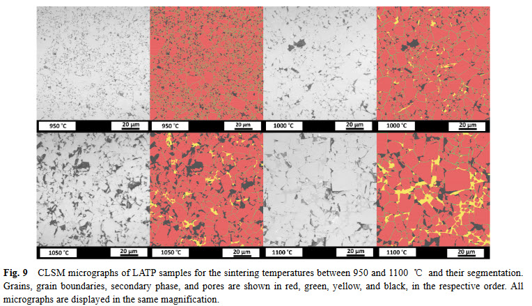
Lithium aluminum titanium phosphate (LATP) is one of the materials under consideration as an electrolyte in future all-solid-state lithium-ion batteries. In ceramic processing, the presence of secondary phases and porosity play an important role. In a presence of more than one secondary phase and pores, image analysis must tackle the difficulties about distinguishing between these microstructural features. In this study, we study the phase evolution of LATP ceramics sintered at temperatures b... Read more
Deniz Cihan GUNDUZ, Roland SCHIERHOLZ, Shicheng YUa, Hermann TEMPEL, Hans KUNGL, Rüdiger-A. EICHEL
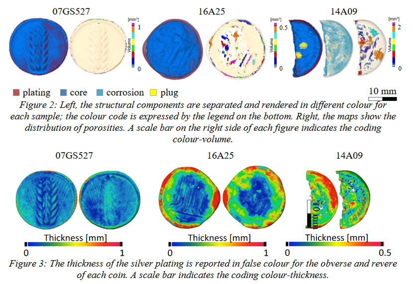
A Neutron Tomographic Analysis of Plated Silver Coins from Ancient Greece Official or Illegal?
In the 6th century BC different techniques of coin manufacture were employed by mints in mainland Greece and in the Greek colonies in Southern Italy. In Greece these techniques were evidently derived from the Lydians and consisted in striking a piece of cast metal of predetermined weight (a ‘blank’ or flan) between two engraved dies made of hardened bronze. Colonies in Magna Graecia, however, uniquely developed another set of minting techniques to produce what today is called incuse coina... Read more
Scott Olsen, Filomena Silvemini, Ulf Garbe, Max Avdeev, Joel Davis, Vladimir Luzin, Ken Sheedy
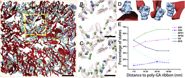
In Situ Structure of Neuronal C9orf72 Poly-GA Aggregates Reveals Proteasome Recruitment
Protein aggregation and dysfunction of the ubiquitin-proteasome system are hallmarks of many neurodegenerative diseases. Here, we address the elusive link between these phenomena by employing cryo-electron tomography to dissect the molecular architecture of protein aggregates within intact neurons at high resolution. We focus on the poly-Gly-Ala (poly-GA) aggregates resulting from aberrant translation of an expanded GGGGCC repeat in C9orf72, the most common genetic cause of amyotrophic latera... Read more
Qiang Guo, Carina Lehmer, Antonio Martinez-Sanchez, Till Rudack, Florian Beck, Hannelore Hartmann, Manuela Perez-Berlanga, Frederic Frottin, Mark S.Hipp, F. Ulrich Hartl, Dieter Edbauer, Wolfgang Baumeister, Ruben Fernandez-Busnadiego
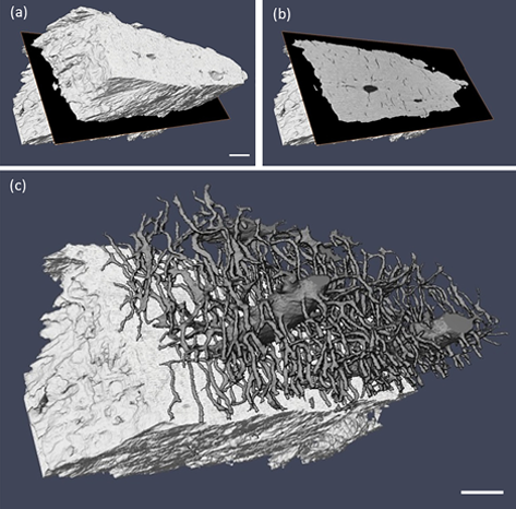
Ptychographic X-ray computed tomography (PXCT) is a quantitative imaging modality that non-destructively maps the 3D electron density inside an object with tens of nanometers spatial resolution. This method provides unique access to the morphology and structure of the osteocyte lacuno-canalicular network (LCN) and nanoscale density of the tissue in the vicinity of an osteocyte lacuna. Our findings indicate that PXCT can non-destructively provide detailed, nanoscale information on the 3D org... Read more
Antonia Ciani, Hechmi Toumi, Stéphane Pallu, Esther H.R.Tsai, Ana Diaz, Manuel Guizar-Sicairos, Mirko Holler, Eric Lespessailles, Cameron M.Kewish | Synchrotron Soleil, France